how do they x ray babies hips
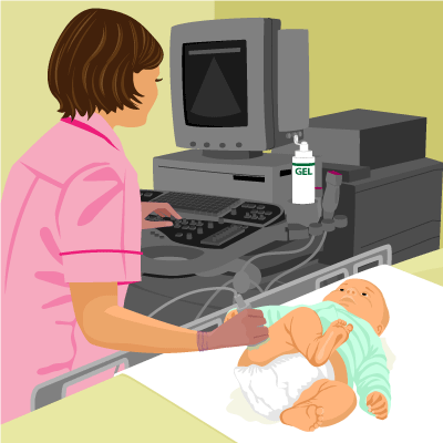
An ultrasound machine sends sound waves into the hip area and images are recorded on a computer. Calcium in your bones takes in more radiation so your bones appear white on the X-ray.
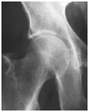
Developmental Dysplasia Of The Hip Prof Portinaro Orthopedic Suregon
How do they x ray babies uk are a topic that is being searched for and liked by netizens today.

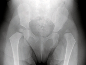
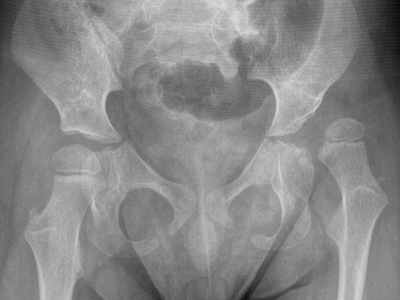
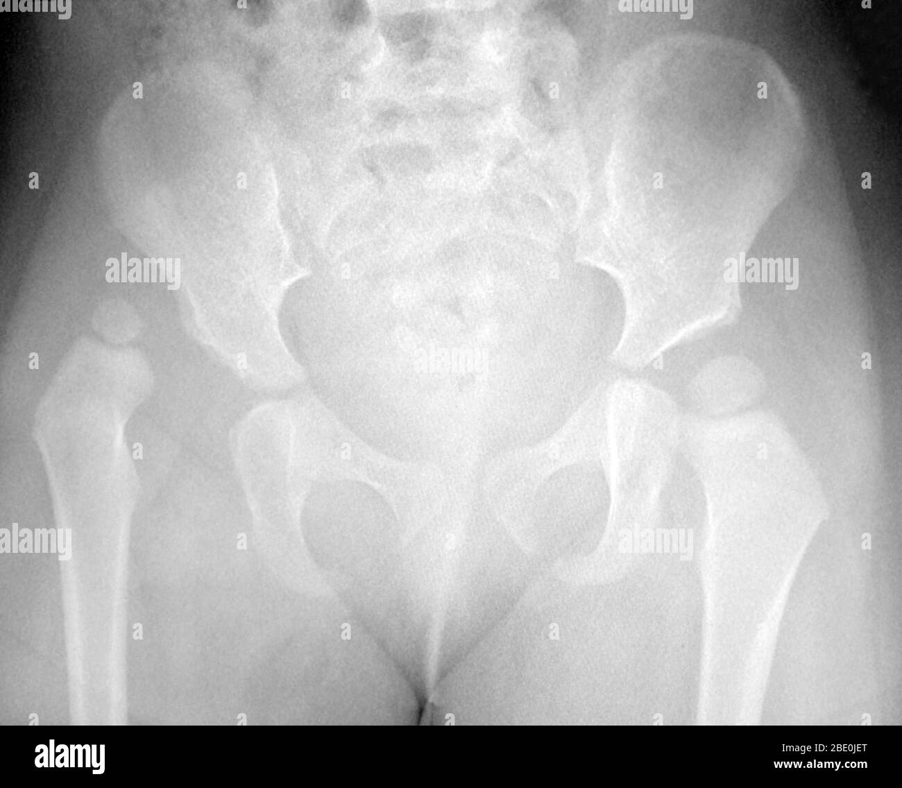
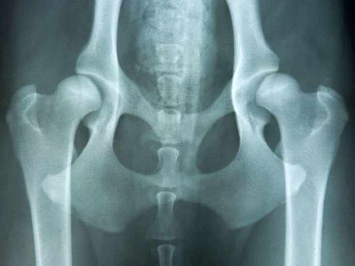
. How do they XRAY a baby hips. The picture shows the inner structure anatomy of your hips in black and white. A hip X-ray radiograph is a medical imaging test that creates a picture of your hip joints and pelvic bones.
Its used because babies and toddlers are incapable of following directions to hold still. A hip X-ray radiograph is a medical imaging test that creates a picture. The main pathology is congenital dislocation of the.
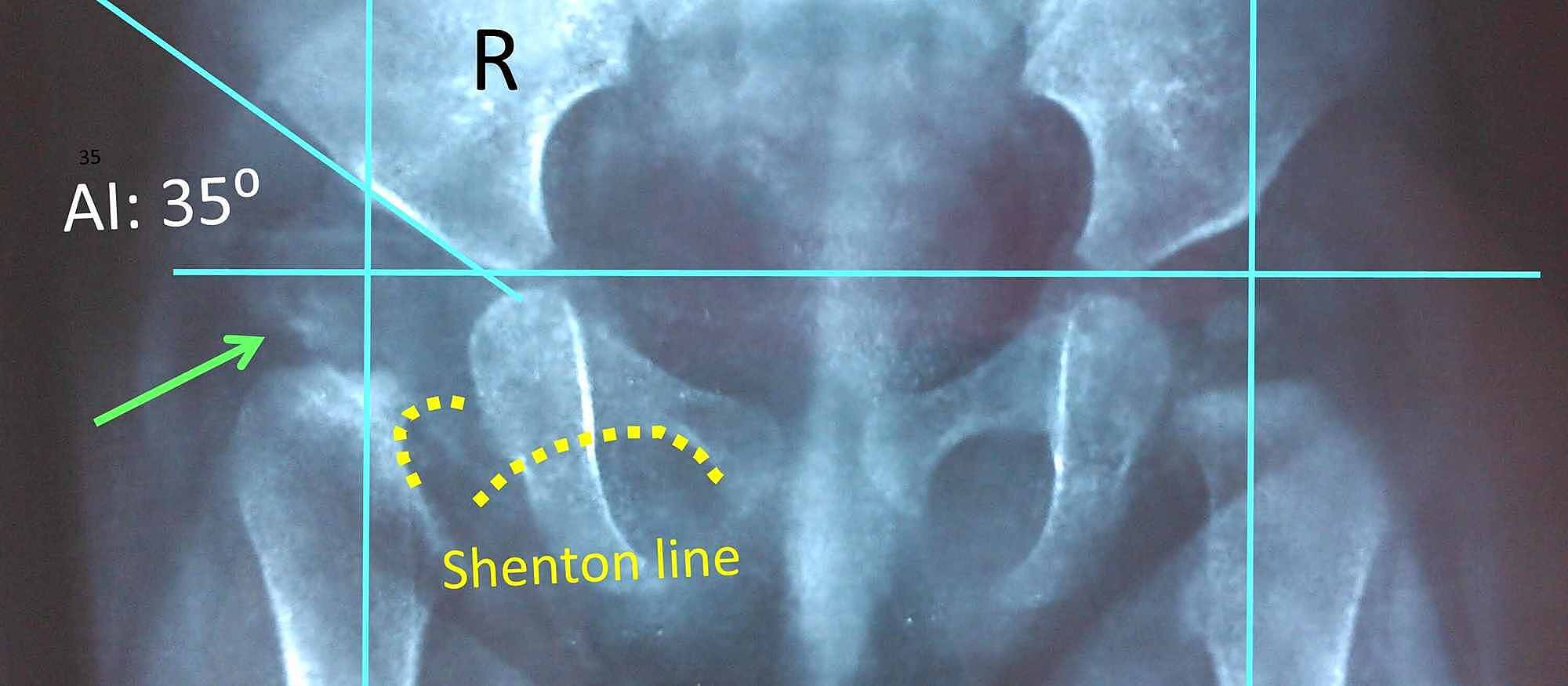
Two tests are performed called the Barlow. It s sometimes called congenital. An X-ray of the pelvis focuses specifically on the area between your hips that holds many of your reproductive and digestive organs.
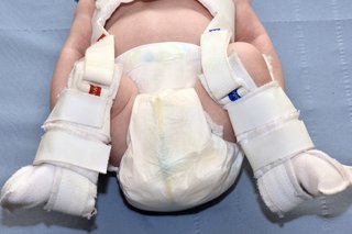
He may also be wheezy after a milky burp. Its a cast that goes around both hips and down the leg to keep the hips. You will go in the room with him he will need to be stripped from the waist down they will take x-rays of him flat on his back legs dead straight.
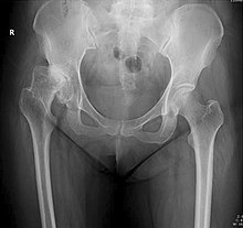
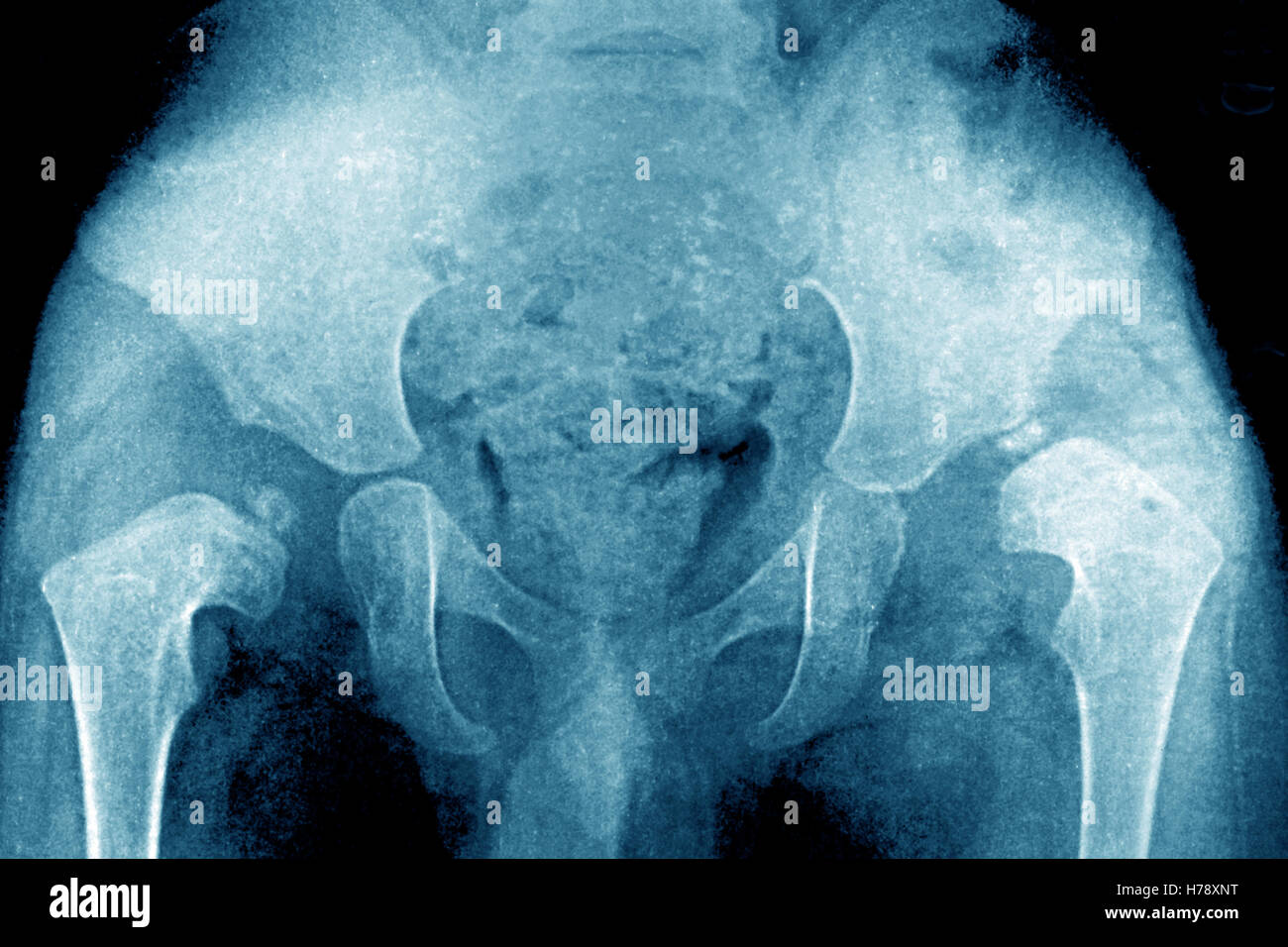
If an X-ray of the hip joints is performed according to Launstein Lauenstein then the patients position looks like this. An X-ray technician will take pictures of the hip. An ultrasound machine sends sound waves into the hip area and images are recorded on a computer.
From the front anteroposterior view or AP from the side lateral view also known as the frog leg lateral view Typically X-rays of both hips are. If she does have it they may try to brace it first. In a hip X-ray an X-ray machine sends a beam of radiation through the pelvic bones and hip joints where the legs attach to the pelvis and an.
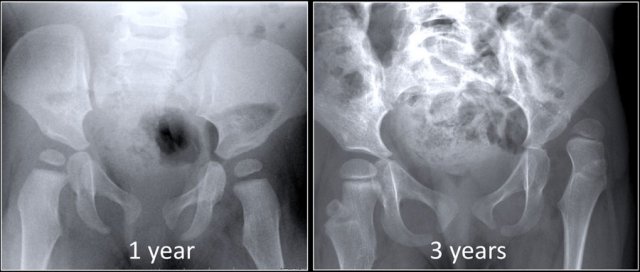
Value How Do They X Ray Babies Hips 2022. The black-and-white images show the internal structures of the hip including the. Around 6 months of age enough bone is present in an infant hip to make an X-ray more accurate than.
X-rays of the hip can reveal bone tumors and diagnose bone cancer. How do they x ray babies hips. Priority How Do They X-Ray Babies Uk 2022.
Totaleclipse 07092007 1731. So far their hips have been fine. An X-ray of the hip joints in children is carried out according to strict indications - only after the child reaches nine months.
Your How do they x ray babies uk images are available. Your How do they. Its a very effective way of looking at the bones and can be used to help detect a range of.
An X-ray of the hip joints in children is carried out according to strict indications - only after the child reaches nine months. If it persists they may put on a spica cast.
X Ray Of Hip Dysplasia Wikipedia

Ce4rt Guide For X Ray Techs To Immobilize Pediatrict Patients
Cureus Instability Testing For Congenital Hip Dislocation Knee Extension Provokes Hip Dislocation

Hip Dysplasia Boston Children S Hospital
Hip Surveillance Helps Identify Dislocations In Children With Cerebral Palsy Children S National

Mackie Orthopaedics Common Paediatric Conditions
X Ray Showing A Dislocated Right Hip On Left Of Image Of A Child Stock Photo Alamy

Xray Hip Child Stock Photo 238386826 Shutterstock
Hip Dislocation Baby Hi Res Stock Photography And Images Alamy
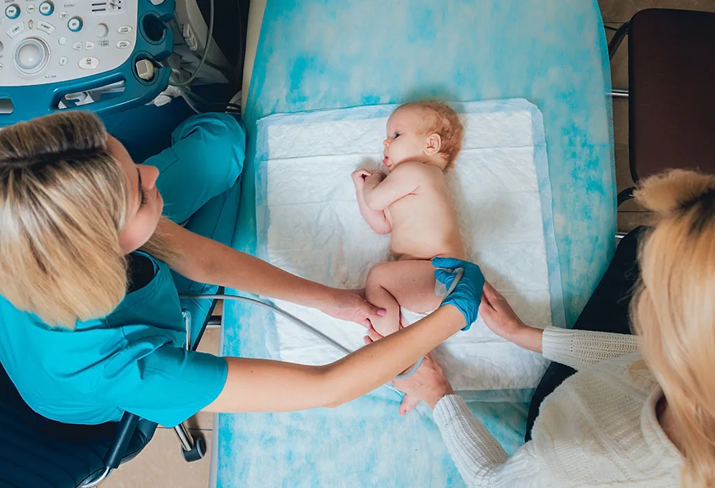
Ultrasound Infant Hip For Parents Nemours Kidshealth

Hip I Developmental Dysplasia Of The Hip Musculoskeletal Key
Developmental Dysplasia Of The Hip Nhs
Hip Dysplasia Information Symptoms Diagnosis Treatment

Child S Pelvis X Ray Stock Image P116 0843 Science Photo Library
Adolescent Hip Dysplasia Orthoinfo Aaos
Hip Dysplasia In Infants Causes Signs Diagnosis Treatment

Hip Dysplasia Boston Children S Hospital
Congenital Hip Dislocation Causes Symptoms And Diagnosis
The Radiology Assistant Developmental Dysplasia Of The Hip